ナノビーム(X線非線形光学)
X線非線形光学現象の研究や、XFEL照射によるダメージ過程の研究には、約100 nmにまでXFELを集光したナノビームが広く利用されています。本ページでは、SACLAにおけるナノビーム実験について紹介します。
なお、EH4cにおいてはより小さなSub-10 nmサイズまで集光したビームが使用可能です。このビームの利用基盤は非常に特殊ですので、詳しくはこのメールアドレスはスパムボットから保護されています。閲覧するにはJavaScriptを有効にする必要があります。までお問い合わせください。
参考文献:
H. Yumoto et al., Appl. Sci. 10, 2611 (2020).
J. Yamada et al., Nat. Photon. 18, 685 (2024)
基本性能・成果
可飽和吸収現象の観測
XFELナノビームにより達成される超高強度X線により、多くの1s軌道電子がXFELのパルス幅内で剥ぎ取られ、X線の透過率が大幅に増加する可飽和吸収現象が、硬X線領域において初めて観測されました。
参考文献:
H. Yoneda et al., Nat. Commun. 5, 5080 (2014).
 Observation of saturable absorption in Fe
X線ダメージ過程の時間分解測定
XFELナノビーム照射による試料のダメージ過程がフェムト秒スケールで観測されました。本実験は、ナノビームと2色XFELとを組み合わせて実施されました。
参考文献:
I. Inoue et al., Proc. Natl. Acad. Sci. USA 113, 1492 (2016).

Time-resolved study of X-ray induced radiation damage on diamond
実験装置
ナノ集光光学系 ‘100exa’
SACLA BL3のEH5には、超高精度に作製された2枚の全反射楕円ミラーで構成された、Kirkpatric–Baez(KB)型のナノ集光光学系が備えられています。典型的な集光サイズは、半値全幅で約200 nm(縦) × 120 nm(横)となります。アンジュレータ出口から集光点までのスループットの目安は約25%であり、集光点でのX線強度は1020 W/cm2(100 EW/cm2)に達するため、本光学系は’100exa’と呼ばれています。全反射ミラーを利用しているため、XFELのエネルギーは12 keVまで色収差の影響を受けること無く利用でき、2色発振XFELを利用した測定も可能です。
本光学系の駆動系は高剛性な石定盤に固定されています。また、真空チャンバーは石定盤とは独立して床面に固定されているため、真空引きによる振動の影響は極めて小さくなります。小型の集光ミラーを利用し、短い焦点距離をとるよう設計されていますので、長期安定性が高く、ナノ集光を12時間以上に渡って安定的に供給可能です。
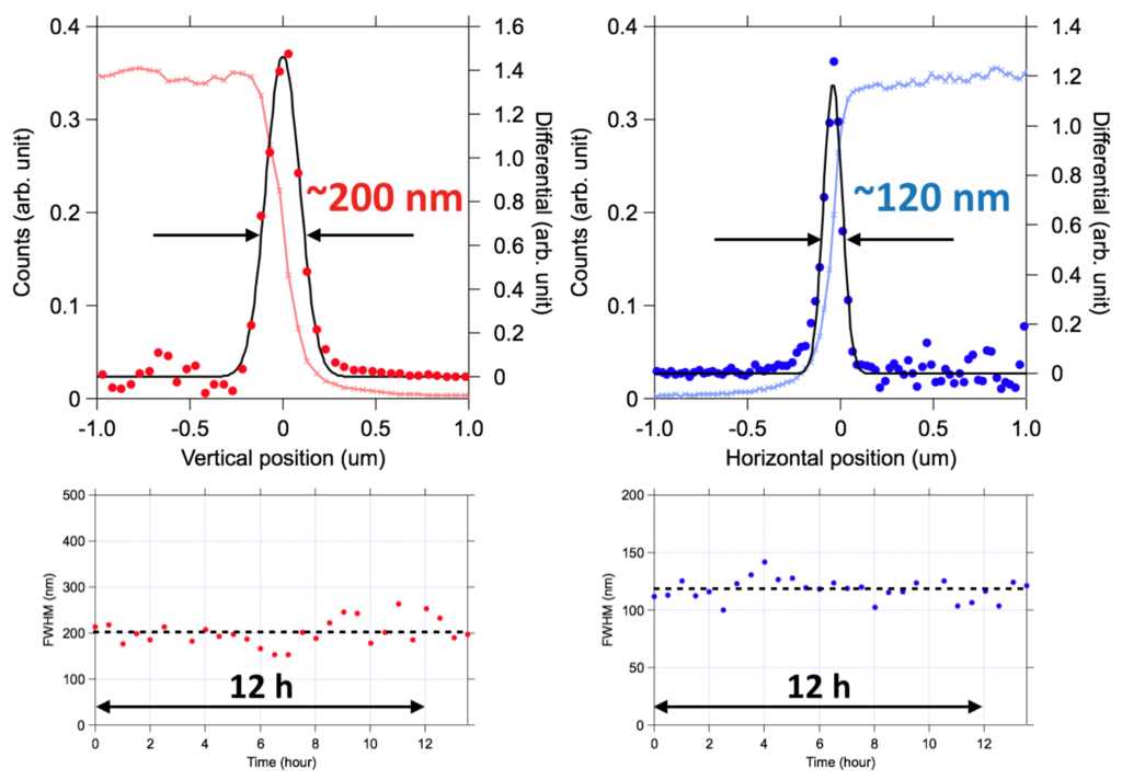
Typical focal profiles measured by knife-edge scanning method (top) and results of stability test (bottom).
Table 1 Optical parameters for 100exa focusing system
| Optical parameters (KB Mirrors) | Vertical | Horizontal |
| Surface coating | Rhodium | Rhodium |
| Substrate size | 250 × 50 × 50 mm | 250 × 50 × 50 mm |
| Glancing angle | 4.0 mrad | 3.8 mrad |
| Focal length | 500 mm | 240 mm |
| Distance from source | 220 m | 220.26 m |
| Effective mirror length |
242 mm | 242 mm |
| Spatial acceptance | 970 μm | 920 μm |
| Divergent angle | ~2 mrad | ~4.5 mrad |
| Typical focal size @10keV | ~200 nm FWHM | ~120 nm FWHM |
参考文献:
H. Yumoto et al., Appl. Sci. 10, 2611 (2020).
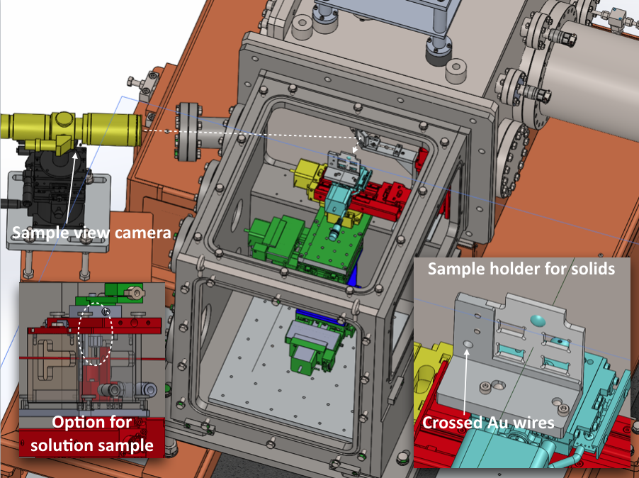
サンプルチャンバー
厚さ75 μmのBe窓を介して100exa光学系真空チャンバーと直接固定が可能なサンプル用真空チャンバーが用意されており、一般的なナノ集光XFEL実験に利用可能です。サンプルチャンバーの最上流部にはX線用の開口が付いた光学ミラーが設置されており、X線とほぼ同軸に上流側からサンプルを観察することが出来ます。この試料モニタの焦点深度は100~200 μmであるため、このモニタを用いることで光軸方向の試料位置を大まかに調整可能です。サンプルチャンバーの上面、下流面、光源を背にして左側面は取替可能で、X線回折や小角散乱、発光分光等、多岐に渡る測定手法に対応できます。また、He等でチャンバー内を置換し、専用の小型ケージを利用することで、溶液サンプルを用いて実験することも可能です。さらに、サンプルチャンバーは取り外し可能ですので、チャンバー付きではアクセスできない位置に検出器や各種測定機器を配置することができます。チャンバーを取り外した際でも、100exaチャンバー下流面に専用のアタッチメントを取り付けることで、サンプル観察用開口付ミラーを設置することが出来ます。サンプル位置・姿勢調整用としてX(光軸垂直面水平方向)、Y(光軸方向)、Z(鉛直方向)、θx(X軸を中心とする回転、入射角に相当)、θz軸の自動ステージを搭載しています。また、高強度XFELを固体試料に照射するとシングルショットで試料が破壊されます。常にフレッシュな領域にXFELを照射するための試料走査用ステージ(X、Z軸)も搭載されています。これらの駆動系は、100exa光学系の駆動系と同じ石定盤に固定されており、前述のサンプルチャンバーとは独立しています。なお、ステージ構成はスペースの許す限り変更可能です。
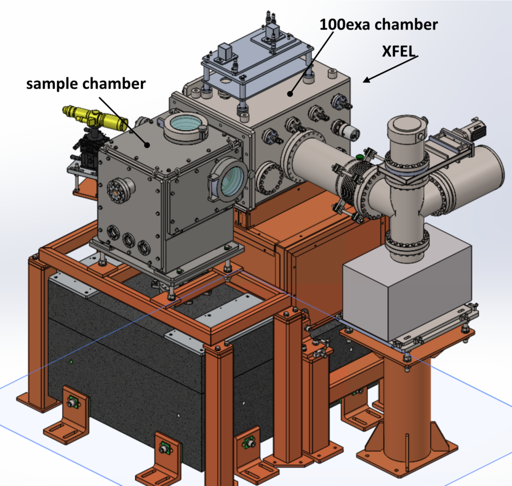
Experimental system with 100exa focusing system.

Schematic layout of sample chamber and control system.
運転パラメータ(EH5@BL3)
ナノビームを使った実験は主にBL3のEH5で行っています。BL3の通常の運転モードである自己増幅自発放射方式(Self-Amplified Spontaneous Emission : SASE)によるXFELに加え、2色XFELやセルフシードXFELを使った実験も可能です。
XFELパラメータ
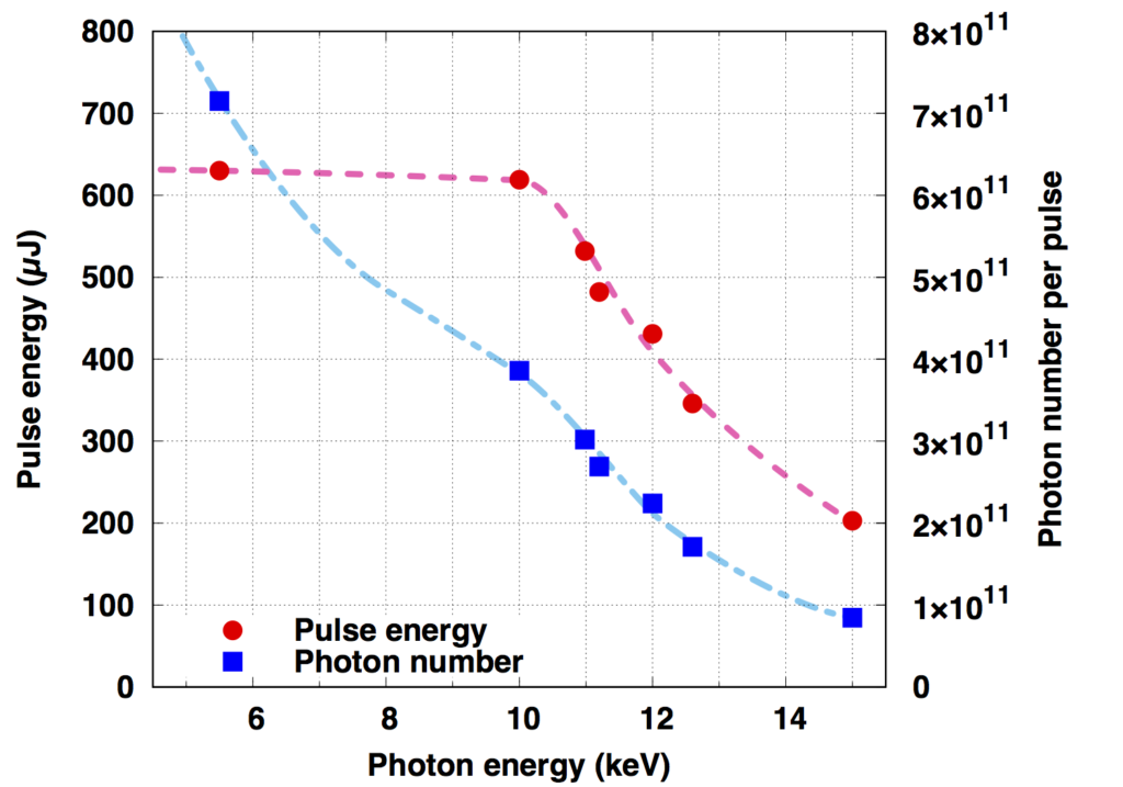
| 光子エネルギー(基本波) | 4-20 keV |
| パルスエネルギー | 光子エネルギーに依存(下図参照) |
| エネルギー幅(ΔE/E) | ~0.5%(ニ結晶分光器なし) |
| 繰り返しレート | 30 Hz(BL2&3同時運転時) |
参考文献:
M. Yabashi et al., J. Synchrotron Rad. 22, 477 (2015).
K. Tono et al., J. Synchrotron Rad. 26, 595 (2019).
(参考)光子エネルギーとパルスエネルギー・光子数の関係(BL3の場合)
2色XFEL
SACLAのアンジュレータ列を2つの区間に分けて、それぞれに異なるギャップを設定することで2種類の波長のXFELを発振させることができます。BL3においては、 1色目のレーザー発振を終えた電子ビームをマグネティックシケインにより迂回させ、1色目と2色目のXFELパルスの間に時間差を付けることが可能です。 遅延時間は数十アト秒の精度で設定することができ、精密なポンププローブ計測などへの応用が期待できます。詳しくはこちらをご覧ください。
参考文献:
T. Hara et al., Nat. Commun. 4, 2919 (2013).
2色発振時のXFELビームパラメーターの例
| 第1色 | 第2色 | |
| 光子エネルギー | 13.1 keV | 9.7 keV |
| パルスエネルギー | 40 µJ | 40 µJ |
| 最大遅延時間 | – | 300 fs |
| 第1色 | 第2色 | |
| 光子エネルギー | 12.2 keV | 9.7 keV |
| パルスエネルギー | 50 µJ | 30 µJ |
| 最大遅延時間 | – | 300 fs |
| 第1色 | 第2色 | |
| 光子エネルギー | 6.1 keV | 5.9 keV |
| パルスエネルギー | 60 µJ | 60 µJ |
| 最大遅延時間 | – | 450 fs |
セルフシードXFEL
BL3において、反射型セルフシード方式によるXFELの利用が可能です。セルフシードXFELは、SASE型XFELと同等の平均パルスエネルギーを有しつつ、高い単色性を示します。詳しくはこちらをご覧ください。
参考文献:
I. Inoue et al., Nature Photon. 13, 319 (2019).
光源パラメーター
| 光子エネルギー | 7 keV-15 keV |
| エネルギー幅 | 0.01% |
| パルスエネルギー | 200 µJ@7-10 keV, ~100 µJ@15 keV |
測定までの調整作業
ビームタイム前までにスタッフが行うアライメント
以下の作業はビームタイム前にSACLAスタッフが行います。
- ・装置の搬入、構築
- ・XFELのスペクトル、光軸、集光の調整
- ・試料観察用カメラの位置、フォーカス調整
ビームタイム開始後にユーザー自身が行うアライメント
ナノビーム実験を行うために、ユーザー自身に行っていただく、最低限のアライメントを以下に示します。
- ・試料位置の調整(ラスタースキャン範囲、光軸方向の位置※)
- ・ユーザー持ち込みの測定系、制御系の調整
※調整方法に関しては、事前にXFEL利用研究推進室までご相談ください。通常、試料からの信号(可飽和吸収等の非線形現象等)をもとに調整します。
ビームタイム開始後にスタッフが行うアライメント
ユーザーからの要望に基づき、必要に応じて100exa光学系の再調整を定期的に行います(ただし9:00~16:30に限る)。
Nanobeam (X-ray Nonlinear Optics)
Nanobeams that focus XFEL to approximately 100 nm are widely used for research on X-ray nonlinear optical phenomenon and damage processes caused by XFEL irradiation. This page will introduce nanobeam experiments performed at SACLA.
References:
H. Yumoto et al., Appl. Sci. 10, 2611 (2020).
J. Yamada et al., Nat. Photon. 18, 685 (2024)
Basic Performance and Results
To observe the saturable absorption phenomenon
For the first time, the saturated absorption phenomenon was observed in the hard X-ray region. This is performed by stripping away many of the 1s orbital electrons within the pulse width of the XFEL with ultra-high intensity X-rays originating from XFEL nanobeams, which greatly increase the transmittance of the X-rays.
References:
H. Yoneda et al., Nat. Commun. 5, 5080 (2014).
Observation of saturable absorption in Fe
Time-resolved measurements of the X-ray damage processes
The damage process of XFEL nanobeam irradiation on the sample was observed on the femtosecond scale. This experiment was carried out in combination with nanobeam and two-color XFEL.
References:
I. Inoue et al., Proc. Natl. Acad. Sci. USA 113, 1492 (2016).
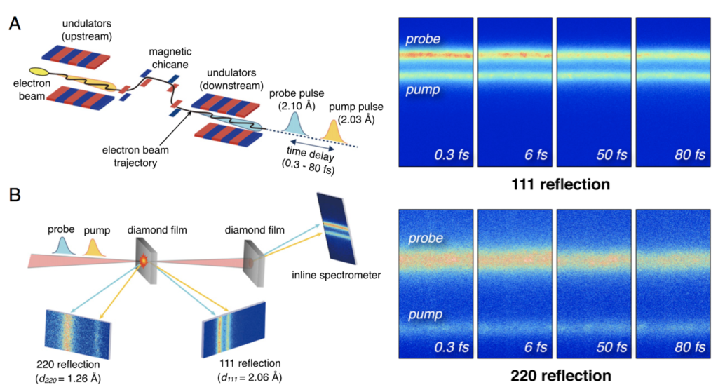
Time-resolved study of X-ray induced radiation damage on diamond
Experimental Equipment
‘100exa’
EH5 at SACLA BL3 features a Kirkpatric-Baez(KB) nano-focusing optical system consisting two ultra-precise total reflection elliptical mirrors. The typical focusing size is approximately 200nm (vertical) and 120nm (horizontal) at full width half maximum. This optical system is called “100exa”, and the approximate throughput from the undulator outlet to the focusing point is approximately 25%, and the X-ray intensity reaches 1020 W/cm2(100 EW/cm2). Since a total reflection mirror is used, the energy of XFEL can be used without being affected by chromatic aberration up to 12 keV, and measurements using two-color oscillations of XFEL is also possible. The driving force for this optical system is fixed to a high-rigidity stone surface plate. In addition, since the vacuum chamber is fixed to the floor surface independently of the stone surface plate, the effects from the vibrations due to vacuuming is extremely small. Designed to use a short focal length using a small focusing mirror, it has high long-term stability that can stably supply nano-focused light for 12 hours or more.

Typical focal profiles measured by knife-edge scanning method (top) and results of stability test (bottom).
Table 1 Optical parameters for 100exa focusing system
| Optical parameters (KB Mirrors) | Vertical | Horizontal |
| Surface coating | Rhodium | Rhodium |
| Substrate size | 250 × 50 × 50 mm | 250 × 50 × 50 mm |
| Glancing angle | 4.0 mrad | 3.8 mrad |
| Focal length | 500 mm | 240 mm |
| Distance from source | 220 m | 220.26 m |
| Effective mirror length |
242 mm | 242 mm |
| Spatial acceptance | 970 μm | 920 μm |
| Divergent angle | ~2 mrad | ~4.5 mrad |
| Typical focal size @10keV | ~200 nm FWHM | ~120 nm FWHM |
References:
H. Yumoto et al., Appl. Sci. 10, 2611 (2020).
Sample chamber
A 100exa optical vacuum chamber, and a sample vacuum chamber with a 75 μm-thick Be window are available for samples that can be directly fixed for general nano-focusing XFEL experiments. An optical mirror with an opening for X-rays is installed upstream of the sample chamber, and the sample can be observed upstream almost coaxially with the X-rays. Since the depth of focus of the sample monitor is 100 to 200 μm, the sample position on the optical axis can be roughly adjusted using this monitor. The upper surface, downstream surface and left surface of the sample chamber can be replaced with a backlit light source, enabling a wide range of measurement methods such as X-ray diffraction, small-angle scattering, and emission spectrometry. It is also possible to experiment with solution samples by filling the chamber with He, or the like, by using a small cage. In addition, the sample chamber is removable, which allows the detector and various other measuring equipment to be placed in locations that are not accessible with the chamber in place. Even when the chamber is removed, a mirror with an opening for the sample observation can be installed by attaching a mount downstream to the 100exa chamber. For sample positioning and orientation adjustments, it is equipped with automatic stages for X (horizontal direction, perpendicular to the optical axis), Y (in the direction of the optical axis), Z (vertical direction) and θx (rotation around the X axis, corresponding to the angle of incidence). In addition, when a solid sample is irradiated with high-intensity XFEL, the sample is destroyed in a single shot. A sample scanning stage (X, Z axis) is also installed to irradiate a fresh area with XFEL at all times. These driving systems are fixed to the same stone surface as the driving system of the 100exa optical system and are independent to the sample chamber described above. The stage configuration can be changed as much as the space allows.

Experimental system with 100exa focusing system.
References:
H. Yumoto et al., Appl. Sci. 10, 2611 (2020).
Operating Parameters (EH5@BL3)
Experiments using nanobeams are generally conducted with EH5 of BL3. In addition to XFEL using the Self-Amplified Spontaneous Emission method (SASE), which is the normal operation mode of BL3, experiments using two-color XFEL and self-seeded XFEL are also possible.
XFEL parameters
| Photon energy (fundamental wave) | 4-20 keV |
| Pulse energy | To photon energy (see figure below) |
| Energy width(ΔE/E) | ~0.5%(without a two crystal spectrometer) |
| Repetition rate | 30 Hz(BL2&3 simultaneous operation) |
References:
M. Yabashi et al., J. Synchrotron Rad. 22, 477 (2015).
K. Tono et al., J. Synchrotron Rad. 26, 595 (2019).
(Reference) The relationship between photon energy and pulse energy / photon number (for BL3)

2-Color XFEL
The undulator column at SACLA can be divided into two intervals, each with a different gap to oscillate XFEL at two wavelengths. In BL3, after the electron beam is finished the laser oscillation of the first color, it is diverted by magnetic chicane, when enables a time difference between the first color and second color XFEL pulses. The delay time can be set with an accuracy of tens of attoseconds and is expected to be applied to precise pump-probe measurements. For more information, please click here.
References:
T. Hara et al., Nat. Commun. 4, 2919 (2013).
Examples of XFEL beam parameters during two-color oscillations
| Color 1 | Color 2 | |
| Photon energy | 13.1 keV | 9.7 keV |
| Pulse energy | 40 µJ | 40 µJ |
| Maximum delay time | – | 300 fs |
| Color 1 | Color 2 | |
| Photon energy | 12.2 keV | 9.7 keV |
| Pulse energy | 50 µJ | 30 µJ |
| Maximum delay time | – | 300 fs |
| Color 1 | Color 2 | |
| Photon energy | 6.1 keV | 5.9 keV |
| Pulse energy | 60 µJ | 60 µJ |
| Maximum delay time | – | 450 fs |
Self-Seed XFEL
At BL3, it is possible to use XFEL by the reflective self-seed method. Self-seed XFEL has the same average pulse energy as the SASE method XFEL, while showing high-monochromaticity. Click here for more information.
References:
I. Inoue et al., Nature Photon. 13, 319 (2019).
| Photon energy | 7 keV-15 keV |
| Energy width | 0.01% |
| Pulse energy | 200 µJ@7-10 keV, ~100 µJ@15 keV |
Adjustments before the Measurement
Alignments performed by the staff before the beam time:
The following tasks are performed by SACLA staff before the beam time.
- ・Bring in and construct the equipment
- ・Adjust and focus the XFEL spectrum and optical axis
- ・Position and focus adjustments of the sample observation camera
Alignments performed by the user after the start of the beam time:
The minimum alignments required by the user to perform the nanobeam experiments are shown below.
- ・Adjust the sample position (raster scan range, *position in the direction of the optical axis)
- ・Adjust the user provided equipment and control systems
※ Please contact the XFEL Utilization Division in advance for adjustment methods. Generally, the adjustments are made based on the signal from the sample (nonlinear phenomenon such as saturated absorption).
Alignments performed by the staff after the start of the beam time:
Based on the user’s request, the 100exa optical system can be readjusted regularly as necessary (only from 9:00 to 16:30).