高角度分解能X線回折装置
◆装置概要
薄膜や表面界面の精密構造解析を行うことができる四軸回折計を基本デザインとした擬似六軸回折装置です。数ミクロンサイズまで集光したX線ビームも利用できます。

◆装置の特徴
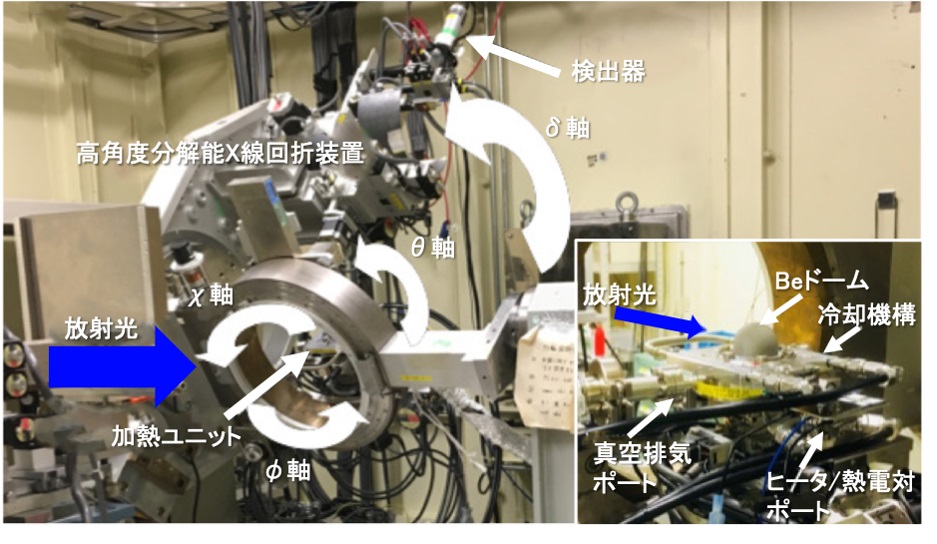
多軸回折計(Kohzu-Seiki TDT-17) を用いて、薄膜構造解析や薄膜と基板との界面構造が解析できます。制御ソフトウェアにはspec(https://www.certif.com)を利用しています。この回折計は標準4軸型のほか、限定的ながら5軸型、6軸型としても使用可能です。4軸型の場合、サンプル周りに3つの自由度(ω軸、χ軸、φ軸)を持ち、検出器には1つの自由度(δ軸すなわち、2θアーム)が充てられています。検出器を設置する2θアームには水平及び回転駆動のステージ(γ軸)があり、これによって、検出器の設置軸は2つの自由度をもち、5軸型の回折計となります。2θアーム上には分光結晶を設置できるので、よりバックグランドを低減し S/N比を改善した測定も実施できます。微小角入射配置での入射角調整軸として利用される回折計全体の回転軸(α軸)を加えれば、6軸型回折計となります。本回折計の各軸の最小分解能を表に示します。
ダブルスリット配置の他に、検出X線のS/N比を向上させる受入れ角0.4度のソーラスリット(Huber 3030-I)も利用できます。検出器には、イオンチャンバ、シリコンPINフォトダイオード検出器、シンチレーション検出器(Oxford x2000)、シリコンドリフト検出器、イメージングプレート検出器、一光子計測二次元検出器(Pilatus-100K, HyPix-400)が利用可能です。
エネルギー領域6 keVから30 keV程度(条件により50 keVまで)のX線が利用でき、最大ビーム強度は6×10¹³ 光子数/秒(@12.4 keV)です。利用時のビームサイズは100 μm程度が標準的ですが、X線屈折レンズを利用した集光光学系を用いれば、数 μmサイズのビームも利用可能です。
【表】 多軸回折計の各軸の最小分解能
| Axis | Step (arc sec) |
| ω | 0.09 |
| φ | 1.7289 |
| ψ | 1.8 |
| 2θ | 0.72 |
| γ | 3.96 |
| α | 0.144 |
◆装置アクセサリー
標準で試料劣化を防止するヘリウム・窒素フローの試料チャンバを利用できる他、高真空中で550 deg.程度まで加熱可能なベリリウム窓付試料チャンバも利用できます。X線照射位置確認のための光学顕微鏡、必要に応じて数μmサイズのビームを生成するX線屈折レンズが利用できます。装置は試料アライメントのためのモーター駆動調整ステージ5軸(平行移動3軸、あおり2軸)および入射ビーム成形スリットを備えており、ビーム位置安定化システム(MOSTAB)も利用されています。
◆実験・試料準備
試料基板サイズが5~10 mm程度であれば、標準の試料チャンバに取り付けできます。それ以外のサイズでの測定を希望される場合はBL担当者にお問い合わせください。事前の試料準備は特に必要とはしませんが、ビームタイム開始時には、毎回、装置の角度精度を保証するために光源・光学系・回折装置のアライメントを実施しています。
試料調整チャンバを持ち込んでの測定を希望される方は、装置と干渉しないか事前に据え付け調整する必要があります。
◆実験手順・注意事項
制御ソフトウェアとして採用しているspec(https://www.certif.com)は、単純なコマンド入力で目的とする逆格子空間へ容易にアクセスできる他、強力なマクロ機能により煩雑な測定手順を自動化できます。一方で、標準のユーザインターフェイスがCUI(Character User Interface)である点にご注意ください。
試料ステージ面からX線照射位置までの距離は40 mmとなっていますので、試料調整チャンバを持ち込んでの測定を希望される方は、装置との干渉に十分注意する必要があります。
本装置を初めて利用される方、あるいは、これまでとは異なる測定法・測定条件を検討しておられる方は、実験課題を効果的に実施するために課題申請前にかならずBL担当者と打ち合わせをおこなうことを推奨します。
◆問い合わせ先
田尻寛男 このメールアドレスはスパムボットから保護されています。閲覧するにはJavaScriptを有効にする必要があります。
◆代表的な論文リスト
[1] “Effect of Non-Specifically Adsorbed Ions on the Surface Oxidation of Pt(111)”
M. Nakamura, Y. Nakajima, N. Hoshi, H. Tajiri, and O. Sakata,
ChemPhysChem, vol.14, pp.2426-2431 (2013).
DOI: 10.1002/cphc.201300404
[2] “Nanosecond Phase Transition Dynamics in Compressively Strained Epitaxial BiFeO3”
M.P. Cosgriff, P. Chen, S.S. Lee, H.J. Lee, L. Pitike, W. Parker, L. Louis, H. Tajiri, S. Nakhmanson, J.Y. Jo, Z. Chen, L. Chen, and P. Evans,
Adv. Electronic Mater., vol.1, pp1500204-(1-9) (2015).
DOI: 10.1002/aelm.201500204
[3] " Large enhancement of bulk spin polarization by suppressing CoMn anti-sites in Co2Mn(Ge0.75Ga0.25) Heusler alloy thin film”
S. Li, Y.K. Takahashi, Y. Sakuraba, N. Tsuji, H. Tajiri, Y. Miura, J. Chen, T. Furubayashi, and K. Hono,
Appl. Phys. Lett., vol. 108, pp122404-(1-5) (2016) .
DOI: 10.1063/1.4944719
[4] “Air/Liquid Interfacial Nanoassembly of Molecular Building Blocks into Preferentially Oriented Porous Organic Nanosheet Crystals via Hydrogen Bonding”
R. Makiura, K. Tsuchiyama, E. Pohl, K. Prassides, O. Sakata, H. Tajiri, and O. Konovalov,
ACS Nano, vol.11 pp10875-10882 (2017).
DOI: 10.1021/acsnano.7b04447
[5] “Effect of hydrophobic cations on the oxygen reduction reaction on single-crystal platinum electrodes”,
T. Kumeda, H. Tajiri, O. Sakata, N. Hoshi, M. Nakamura,
Nature Commun., vol. 9, 4378-(1-7) (2018).
DOI: 10.1038/s41467-018-06917-4
High-Angular Resolution X-ray Diffraction Equipment
◆Equipment overview
This is a pseudo-six-axis diffractometer with a basic design of a four-axis diffractometer, which can perform precise structural analysis for thin film surfaces. There are also available X-ray beams that are focused to several microns in size.

◆Features of the Equipment
Miltiple-axis diffractometers (Kohzu-Seiki TDT-17) are used for thin film structural analysis and interface structure analysis between thin films and substrates. The control software being used is Spec (https://www.certif.com).This diffractometer can be used as a standard 4-axis type, as well as a limited 5-axis or 6-axis type. In the case of a 4-axis type, there are 3 degrees of freedom (ω-axis,χ-axis,φ-axis) around the sample, and 1 degree of freedom (δ-axis, or 2θ arm) for the detector. The 2θ arm installed on the detector has a level and rotational driving stage (γ-axis), which allows the detector to have 2 degrees of freedom and a 5-axis diffractometer. Since a light-coded crystal can be installed on the 2θ arm, measurements with reduced background and improved S/N ratio can be performed. If you add the rotation axis (α-axis) of the entire diffractometer used as the incident angle adjustment axis in the micro-angle incident arrangement, it becomes a 6-axis diffractometer. The table shows the minimal resolution of each axis of this diffractometer.
In addition to the double-slit arrangement, a 0.4 degree solar-slit (Huber 3030-I) is also available, which improves the S/N ratio of the X-ray detection. The following equipment is available for detectors: an ion chamber, a silicon PIN photodiode detector, a scintillation detector (Oxford x2000), a silicon drift detector, an imaging plate detector, and a single-photon measurement 2-dimensional detector (Pilatus-100K, HyPix-400).
X-rays in the energy region of 6 keV to 30 keV (up to 50 keV depending on the conditions) are available, with a maximum beam intensity of 6×10¹³ photons/second (@12.4 keV). The typical beam size used is 100 μm, but a beam of several μm is also available when using a focusing optical system with a refractive X-ray lens.
Table: Minimum resolution of each axis of the multi-axis diffractometer
| Axis | Step (arc sec) |
| ω | 0.09 |
| φ | 1.7289 |
| ψ | 1.8 |
| 2θ | 0.72 |
| γ | 3.96 |
| α | 0.144 |
◆Equipment accessories
In addition to a Helium-Nitrogen flow sample chamber to prevent sample deterioration, a sample chamber with a beryllium window that can be heated up to 550 degrees in a high vacuum is available. Optical microscopes for X-ray irradiation position review, as well as X-ray refraction lenses for generating beams of several μm sizes are also available if necessary. The device features a motor driven adjustment stage with 5 axes (3 translation axes, 2 impact axes) for sample alignment, as well as an incident beam forming slit, and a beam position stabilization system (MOSTAB).
◆Experiment / sample preparation
If the sample substrate size is between 5~10 mm, a standard sample chamber can be installed. If you intend to take measurements beyond this size range, please contact the person in charge of the BL. Prior sample preparation is not required, but every time, at the start of beam time, light sources, optical equipment and diffraction devices are aligned to ensure the angle accuracy of the device.
If you wish to bring in a sample adjustment chamber for measurements, it is necessary to install and adjust in advance to prevent interference with the device.
◆Experimental procedure / precautions
The control software used is Spec (https://www.certif.com), which allows for simple command input that provides easy access to the reciprocal lattice space, where powerful macro functions automate troublesome measurement procedures. On the other hand, please note that the standard user interface is the CUI (Character User Interface).
Since the distance of the sample stage surface to the X-ray irradiation position is 40 mm, if you wish to bring a sample adjustment chamber for measurements, pay close attention to interference with the device.
If you are using this device for the first time, or if you are considering different measurement methods or conditions, in order to effectively implement the experimental objectives, it is recommended that you meet with the person in charge of the BL before applying for the assignment.
◆Contact
田尻寛男 このメールアドレスはスパムボットから保護されています。閲覧するにはJavaScriptを有効にする必要があります。
◆List of representative treatises
[1] “Effect of Non-Specifically Adsorbed Ions on the Surface Oxidation of Pt(111)”
M. Nakamura, Y. Nakajima, N. Hoshi, H. Tajiri, and O. Sakata,
ChemPhysChem, vol.14, pp.2426-2431 (2013).
DOI: 10.1002/cphc.201300404
[2] “Nanosecond Phase Transition Dynamics in Compressively Strained Epitaxial BiFeO3”
M.P. Cosgriff, P. Chen, S.S. Lee, H.J. Lee, L. Pitike, W. Parker, L. Louis, H. Tajiri, S. Nakhmanson, J.Y. Jo, Z. Chen, L. Chen, and P. Evans,
Adv. Electronic Mater., vol.1, pp1500204-(1-9) (2015).
DOI: 10.1002/aelm.201500204
[3] " Large enhancement of bulk spin polarization by suppressing CoMn anti-sites in Co2Mn(Ge0.75Ga0.25) Heusler alloy thin film”
S. Li, Y.K. Takahashi, Y. Sakuraba, N. Tsuji, H. Tajiri, Y. Miura, J. Chen, T. Furubayashi, and K. Hono,
Appl. Phys. Lett., vol. 108, pp122404-(1-5) (2016) .
DOI: 10.1063/1.4944719
[4] “Air/Liquid Interfacial Nanoassembly of Molecular Building Blocks into Preferentially Oriented Porous Organic Nanosheet Crystals via Hydrogen Bonding”
R. Makiura, K. Tsuchiyama, E. Pohl, K. Prassides, O. Sakata, H. Tajiri, and O. Konovalov,
ACS Nano, vol.11 pp10875-10882 (2017).
DOI: 10.1021/acsnano.7b04447
[5] “Effect of hydrophobic cations on the oxygen reduction reaction on single-crystal platinum electrodes”,
T. Kumeda, H. Tajiri, O. Sakata, N. Hoshi, M. Nakamura,
Nature Commun., vol. 9, 4378-(1-7) (2018).
DOI: 10.1038/s41467-018-06917-4
