高エネルギーX線構造解析装置
◆装置概要
汎用的に有機、無機物質の高分解能構造解析を行うことができます。機能性材料の電子密度レベルの精密解析などに利用されています。
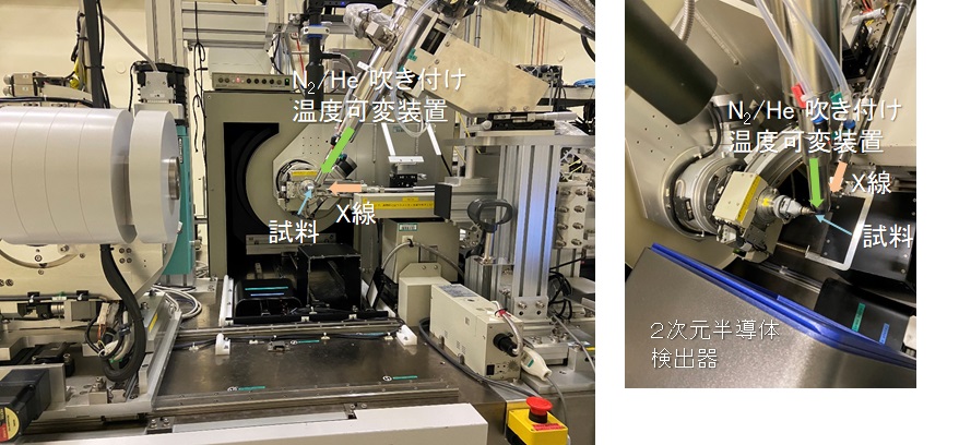
装置写真:高エネルギーX線対応の半導体検出器を用いた単結晶構造解析装置

◆装置の特徴
本装置は、汎用的に有機、無機物質の高分解能構造解析を行うことができます。機能性材料の電子密度レベルの精密解析などに利用されています。1/4χゴニオメータを搭載し、検出器として半導体検出器のPILATUS3 X CdTe 1M及び湾曲型のイメージングプレートを切り替えて使用することが可能です。・1/4χ回折計
ω、χ、φ軸の3軸を搭載した回折計であり、構造解析に必要とされる逆空間を網羅的に観測することが可能です。(IP: 2θmax < 145°; PILATUS: 2θmax < 65°)
・検出器
・高エネルギーX線対応半導体検出器
本検出器は、CdTeを検出素子として採用した高エネルギー対応の半導体検出器です。回折像の高速読み出しが可能であり、短時間で単結晶構造解析データを収集することが可能です。詳しい仕様はこちらから。
・大型湾曲IPカメラ
X線エネルギーの選択性がほとんどなく、広いダイナミックレンジを有し、大面積を一度に観測できる検出器です。主に、電子密度分布などの精密構造解析に使用されます。詳しい仕様はこちらから。
・制御ソフトウェア
実験室の装置でも使用されているリガク社製の制御ソフト「Control」を半導体・大型湾曲IPカメラで使用しており、実験室と同様の操作で回折データの収集が可能です。
◆装置アクセサリー
本システムでは、下記のアクセサリーの使用が可能です。
・ヘリウムガス吹付け低温装置(20〜400K)
・高温窒素ガス吹き付け装置(300〜800K)
・紫外線照射装置
・キセノン光源
・高圧電場印加装置
・X線パルスセレクター
◆実験・試料準備
測定の際に試料を取り付けるホルダは実験室系の装置で一般的に使用されるマイクロマウントやループ、またはガラス針を利用できる(ベースからの高さ20mm程度)。ゴニオメータヘッドにはマグネットやφ3mmの金属棒部分で保持することが可能です。空気中で溶媒脱離などにより結晶性が劣化する場合にはビームラインに設置された実体顕微鏡を利用してビームラインでの試料取り付けも可能です。
◆実験手順・注意事項
単結晶構造解析の実験は、実験室の装置とほぼ同様の手順で行えるようになっています。下記に一般的な単結晶構造解析実験の手順を示します。
1)試料を試料ホルダに取り付ける
試料ホルダは一般的に実験室系で利用されるマイクロマウント、ループ、ガラス針が利用できる。(ベースからの高さ20mm程度)
2)試料ホルダを回折計に取り付ける
3)センタリング
φ、ω軸に対してセンタリングを行います。
4)仮測定による結晶の評価
短時間(数分間)の測定を行って結晶性や回折強度をチェックします。
5)データ測定
チェック測定の結果を基にして露光時間やX線強度を決定してデータ測定を行う。 通常はφ:0,90,180,270°の各位置でω:0~180°の範囲をΔω<1°毎に回折像を取得する。PILATUSの読み取り時間が短いため、シャッターレス測定が行われ、1秒露光の場合約12分で全反射の測定が完了する。
6)回折強度の積分
ビームラインに導入されている積分用ソフトウェアを利用して回折像から回折強度を抽出する。 利用可能なソフト:Rapid Auto、CrysAlisPro(RIGAKU)、 APEX3(Bruker)
7)構造解析
積分処理によって得られた結晶学データおよび回折強度データから構造解析を行う。実験後に大学などで解析を進める場合も多く、実験者の使い慣れたソフトでの解析を行うため、原則として構造解析用のソフトウェアはユーザーが準備することを推奨している。
◆問い合わせ先
杉本邦久(このメールアドレスはスパムボットから保護されています。閲覧するにはJavaScriptを有効にする必要があります。)
安田伸広(このメールアドレスはスパムボットから保護されています。閲覧するにはJavaScriptを有効にする必要があります。)
中村唯我(このメールアドレスはスパムボットから保護されています。閲覧するにはJavaScriptを有効にする必要があります。)
◆代表的な論文リスト
"Observation of the Asphericity of 4f-electron Density and its Relation to the Magnetic Anisotropy Axis in Single-Molecule Magnets"
Gao Chen et al.
Nature Chemistry, 12, (2020) 213-219
DOI : 10.1038/s41557-019-0387-6
"X-ray Electron Density Investigation of Chemical Bonding in van der Waals Materials"
Kasai Hidetaka et al.
Nature Materials, 17, (2018) 249-252
DOI : 10.1038/s41563-017-0012-2
"A Linear Cobalt(II) Complex with Maximal Orbital Angular Momentum from a Non-Aufbau Ground State"
Bunting Philip C. et al.
Science, 362, (2018) eaat7319
DOI : 10.1126/science.aat7319
"Observation of Quadrupole Helix Chirality and its Domain Structure in DyFe3(BO3)4"
Usui T. et al.
Nature Materials, 13, (2014) 611-618
DOI : 10.1038/NMAT3942
"Electronic Nematicity above the Structural and Superconducting Transition in BaFe2(As1-xPx)2"
Kasahara Shigeru et al.
Nature, 486, (2012) 382-385
DOI : 10.1038/nature11178
"Spin-Orbital Short-Range Order on a Honeycomb-Based Lattice"
Nakatsuji Satoru et al.
Science, 336, (2012) 559-563
DOI : 10.1126/science.1212154
"Photoactivation of a Nanoporous Crystal for On-Demand Guest Trapping and Conversion"
Sato Hiroshi et al.
Nature Materials, 9, (2010) 661-666
DOI : 10.1038/NMAT2808
"A Layered Ionic Crystal of Polar Li@C60 Superatoms"
Aoyagi Shinobu et al.
Nature Chemistry, 2, (2010) 678-683
DOI : 10.1038/NCHEM.698
High-Energy X-Ray Structural Analysis Equipment
◆Equipment overview
General-purpose high-resolution structural analysis of organic and inorganic substances can be performed. This can be used for precise analysis of the electron density levels of functional materials.
Equipment Photo: Single crystal structural analysis device using a high-energy X-ray compatible semiconductor detector.

◆Features of the Equipment
This equipment can perform general-purpose, high-resolution structural analysis of organic and inorganic substances. This can be used for precise analysis of the electron density levels of functional materials. Equipped with a 1/4χ goniometer, it is possible to switch between the PILATUS3 X CdTe 1M semiconductor detector and a curved imaging plate.・1/4χ Diffractometer
The diffractometer has three axes, ω, χ, and φ, which allows for comprehensive observation of the reciprocal space required for structural analysis.(IP: 2θmax < 145°; PILATUS: 2θmax < 65°)
・Detector
・High-energy X-ray Compatible Semiconductor Detector
This detector is a high-energy semiconductor detector which uses CdTe as the detection element. It is possible to read diffraction images at high speeds, as well as collect crystal structure analysis data in a very short time. Click here for detailed specifications.
・Large Curvature IP Camera
This detector has little selectivity for X-ray energy, but it has a wide dynamic range, and can observe a large area at once. It is used mainly for precision structure analysis, such as electron density distribution. Click here for detailed specifications.
・Control Software
The control software is manufactured by Rigaku, and is also used in the laboratory equipment, from the semiconductor to the large curvature IP camera, this software makes the collection of diffraction data possible.
◆Equipment accessories
The following accessories can be used with this system.
・Low-temperature Helium gas spray equipment (20 – 400 K)
・High-temperature Nitrogen gas spray equipment (300 – 800 K)
・Ultraviolet Irradiation Device
・Xenon light source
・High-pressure electric field application apparatus
・X-Ray pulse selector
◆Experiment / sample preparation
While taking measurements, laboratory equipment sample holders are available, including micromounts, loops or glass needles (with the height from the base about 20mm). The goniometer head can be held with a magnet or a φ3mm metal rod. When crystallinity deteriorates due to solvent desorption in air, it is possible to mount the sample at the beamline using a stereo microscope installed on the beamline.
◆Experimental procedure / precautions
Experiments on single crystal structure analysis can be performed using the same procedure as the laboratory equipment. The following are the procedures for general single structure crystal analysis experiments.
1)Mount the sample in the sample holder
Sample holders such as micromounts, loops and glass needles are commonly used in laboratory systems. (Height from the base is about 20mm)
2)Mount the sample holder on the diffractometer
3)Centering
Centering is performed on the φ and ω axes.
4)Evaluation of crystals by provisional measurements
The crystallization and diffraction strength are checked by measuring for a short time (several minutes).
5)Data measurement
Based on the outcome of the check measurements, the exposure time and X-ray intensity are decided and data measurements are completed. Generally, diffraction images are obtained every Δω<1° within the range ω:0~180°at the φ:0,90,180,270°positions. The reading time of PILATUS is short, and shutterless measurement is performed. For 1 second exposure, the total reflection measurement is completed in approximately 12 minutes.
6)Diffraction Intensity Integration
Diffraction intensity is extracted from the diffraction image using the integration software introduced in the beamline. The available software is: Rapid Auto, CrysAlisPro(RIGAKU), APEX3(Bruker)
7)Structure Analysis
Structure analysis such as crystallography data as well as diffraction intensity data are obtained from performing integration processing. In general, it is recommended that users prepare software for structural analysis that is familiar to them, as analysis is generally carried out at universities and other facilities after the experiment.
◆Contact
杉本邦久(このメールアドレスはスパムボットから保護されています。閲覧するにはJavaScriptを有効にする必要があります。)
安田伸広(このメールアドレスはスパムボットから保護されています。閲覧するにはJavaScriptを有効にする必要があります。)
中村唯我(このメールアドレスはスパムボットから保護されています。閲覧するにはJavaScriptを有効にする必要があります。)
◆List of representative treatises
"Observation of the Asphericity of 4f-electron Density and its Relation to the Magnetic Anisotropy Axis in Single-Molecule Magnets"
Gao Chen et al.
Nature Chemistry, 12, (2020) 213-219
DOI : 10.1038/s41557-019-0387-6
"X-ray Electron Density Investigation of Chemical Bonding in van der Waals Materials"
Kasai Hidetaka et al.
Nature Materials, 17, (2018) 249-252
DOI : 10.1038/s41563-017-0012-2
"A Linear Cobalt(II) Complex with Maximal Orbital Angular Momentum from a Non-Aufbau Ground State"
Bunting Philip C. et al.
Science, 362, (2018) eaat7319
DOI : 10.1126/science.aat7319
"Observation of Quadrupole Helix Chirality and its Domain Structure in DyFe3(BO3)4"
Usui T. et al.
Nature Materials, 13, (2014) 611-618
DOI : 10.1038/NMAT3942
"Electronic Nematicity above the Structural and Superconducting Transition in BaFe2(As1-xPx)2"
Kasahara Shigeru et al.
Nature, 486, (2012) 382-385
DOI : 10.1038/nature11178
"Spin-Orbital Short-Range Order on a Honeycomb-Based Lattice"
Nakatsuji Satoru et al.
Science, 336, (2012) 559-563
DOI : 10.1126/science.1212154
"Photoactivation of a Nanoporous Crystal for On-Demand Guest Trapping and Conversion"
Sato Hiroshi et al.
Nature Materials, 9, (2010) 661-666
DOI : 10.1038/NMAT2808
"A Layered Ionic Crystal of Polar Li@C60 Superatoms"
Aoyagi Shinobu et al.
Nature Chemistry, 2, (2010) 678-683
DOI : 10.1038/NCHEM.698
