高エネルギーX線イメージング装置
◆装置概要
白色X線に重金属フィルタを組み合わせることによって得られる、約200keVのピークエネルギーを持った高エネルギー白色X線を用いることで、10μm程度の空間分解能で金属試料や化石試料等の3Dイメージングが可能です。
◆装置の特徴
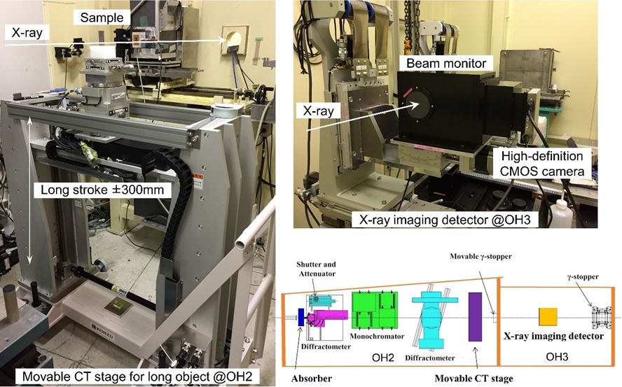
・アブソーバー偏光電磁石からの白色X線から高エネルギーX線成分を取り出します。アブソーバーは0.5mm厚のタングステンプレートと2mm厚の鉛板で構成されます。これにより、約200keVにピークを持った白色X線スペクトルを切り出すことができます。また、アブソーバー自身を高速回転させることにより、アブソーバー由来のスペックルノイズを低減します。
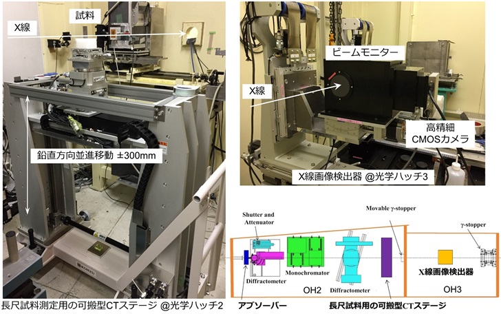
・長尺試料測定用試料ステージ
CT測定を行うための自動ステージ(水平方向並進移動、鉛直方向並進移動、回転軸傾き調整用スイベルステージ2軸、回転ステージ)によって構成されています。特に鉛直方向の自動ステージは、長尺試料の測定を可能とするため、±300mmのストロークを有しています。ステージ全体が可搬型となっており、ハッチ内外への出し入れを行うことが可能です。
・X線画像検出器
可視光変換型のX線画像検出器です。検出器へ入射した高エネルギーX線は、単結晶あるいはセラミクス蛍光体によって可視光へ変換され、タンデムカメラレンズ光学系を介して、高精細CMOSカメラで画像として出力されます。タンデムレンズ系の組み合わせを変えることにより、画素サイズ3μmから12μm程度でのCT測定が可能となります。また、高精細CMOSカメラを用いることにより、広視野かつ高分解能測定の両立が可能となり、最大視野幅50mm程度のCT測定まで対応することが可能です。CMOSカメラは、専用の制御ソフトウエアにより、ライブ観察や外部トリガーによる撮影制御を行うことができます。
◆装置アクセサリー
試料をステージに取り付けるための治具等は、基本的にはユーザーさんご自身により準備していただくことになります。ただし、特殊な形状な試料や、文化財など取り扱いが難しい試料等については、治具の設計に際し必要なアドバイスをさせていただきます。下記の問い合わせ先へご相談ください。また、CT測定用試料ステージは、ユーザーさん側での持ち込み装置への置き換えも可能ですので、ご相談ください。
◆実験・試料準備
高エネルギーX線マイクロCTでは、実効画素サイズ3μmから12μmでの測定になります。このときの撮像視野の幅は、約12mm(画素サイズ3μm時)、約50mm(画素サイズ12μm時)となります。CT測定の特性上、試料サイズ(円柱状の試料を想定した場合のその直径サイズ)は、撮像視野よりも小さいことが望ましいですが、必要な空間分解能に対して、文化財など貴重な試料故に試料サイズの調整が困難な場合も想定されますので、その場合はご相談ください。
実験では、高精細CMOSカメラの利用により、データ容量は非常に大きくなる傾向があります。測定条件にも依りますが、測定データのみで最大4TB/1日のデータ容量となりますので、データ持ち帰り用のストレージディスクは余裕をもってご準備されることをお勧めします。
◆実験手順・注意事項
まず、試料をステージに固定します。
専用の取り付け治具がある場合は、それを用いてください。
取り付けが困難と予想される場合は事前にご相談ください。
長尺試料の場合は、取り付け時の試料の傾きを補正する必要もありますので、その場合は墨だしレーザー等の利用が可能です。
また、専用治具をステージにネジ止めする際に、無理矢理取り付けようとすると、ステージのネジ穴の破損に繋がりますのでご注意ください。
200keVでの高エネルギーX線CT測定では、縦方向の有効視野サイズは1.5mm程度ですので、試料サイズあるいは測定関心領域の大きさによっては、試料を鉛直方向に走査(Zスキャン)する必要があります。この場合、測定開始位置および必要な走査回数(測定長さ)を計測用ソフトウエアに入力します。
測定は、指定した測定情報を元に自動で行います。
通常、Zスキャンを行う前に、測定条件の検討や計測用ソフトウエアの使用方法の習得を兼ねて、走査はせずにCT測定および再構成処理を行い、画像データを確認します。測定条件が決定したら、Zスキャンを伴った本測定を行います。
測定後は、測定したデータのバックアップおよび必要に応じて再構成処理を行います。
再構成処理では、ビームライン保有の高性能計算機の利用が可能です。
◆問い合わせ先
星野真人 このメールアドレスはスパムボットから保護されています。閲覧するにはJavaScriptを有効にする必要があります。
安武正展 このメールアドレスはスパムボットから保護されています。閲覧するにはJavaScriptを有効にする必要があります。
◆代表的な論文リスト
“High-energy, high-resolution x-ray imaging for metallic cultural heritages”
Masato Hoshino, Kentaro Uesugi, Ryuji Shikaku and Naoto Yagi
AIP Advances, 7, (2017) 105122
DOI https://doi.org/10.1063/1.5003162
“Development of an X-ray imaging detector for high-energy X-ray microtomography”
Masato Hoshino, Kentaro Uesugi and Naoto Yagi
Journal of Synchrotron Radiation, 27, (2020) 934-940
DOI https://doi.org/10.1107/S1600577520004920
High-Energy X-Ray Imaging Equipment
◆Equipment overview
It is possible to perform 3-dimensional imaging of metal and fossil samples with a spatial resolution of 10μm. This is done by using high-energy white X-rays with a peak energy of about 200 keV, which are obtained by combining white X-rays with heavy metal filters.
◆Features of the Equipment
・AbsorberHigh-energy X-ray components are extracted from white X-rays by polarized electromagnets. The absorber consists of a 0.5 mm thick tungsten plate and a 2 mm thick lead plate. This allows for white X-rays with a peak of 200 keV to be extracted. In addition, by rotating the absorber at high speeds, the speckle noise which comes from the absorber is reduced.
・Sample Stage for Long Sample Measurements
This system consists of an automatic stage (horizontal and vertical translation movement, 2-axis swivel stage for rotational axis tilt adjustments, and a rotational stage) which is used for performing CT measurements. In particular, the automatic vertical stage has a stroke of ±300mm to enable measurements of long samples. The entire stage is portable, and can be taken in and out of the hatch.
・X-Ray Imaging Detector
The detector is a visible light conversion X-ray imaging detector. High-energy X-rays incident on the detector are converted to visible light by single crystals or ceramic phosphor, then output as an image by a high-definition CMOS camera through tandem camera lens optics. By changing the combination of tandem lens systems, CT measurements are possible with a pixel size of 3 μm to 12 μm. In addition, by using a high-definition CMOS camera, it is possible to achieve both wide field of view and high-resolution measurements. CT measurements can be performed with a maximum field of view of about 50 mm. The CMOS camera can perform live observations and photography control with external triggers using the dedicated control software.
◆Equipment accessories
To begin, the user prepares the jigs to attach the sample to the stage. However, for samples with special shapes or samples that are difficult to handle, we will provide the necessary advice for designing the jigs. In addition, the sample stage for the CT measurements can be replaced with user provided equipment. Please contact us for any inquiries.
◆Experiment / sample preparation
The high-energy X-ray micro-CT measures an effective pixel size of 3 μm to 12 μm. The width of the imaging field within that range is approximately 12 mm (when the pixel size is 3 μm) and approximately 50 mm (when the pixel size is 12 μm). Due to the characteristics of CT measurements, the sample size (the diameter size when assuming a cylindrical sample) should be smaller than the imaging field to achieve the required spatial resolution. If the sample is difficult to adjust, or a valuable sample such as cultural property, please contact us.
During the experiments, the high-definition CMOS camera tends to result in very large volumes of data. It is recommended that you prepare a storage disk to take the data home, as the measurement data alone can be up to 4TB/day depending on the measurement conditions.
◆Experimental procedure / precautions
First, affix the sample to the stage.
Prepare the special mounting jig if you plan to use one.
Please contact us in advance if you expect the sample to be difficult to install.
In the case of a long sample, the inclination of the sample must be corrected when the sample is mounted, and this can be done using ink lasers or the like.
Also, please note that if you try to forcibly attach the special jig to the stage, it may damage the screw holes on the stage.
For high-energy X-ray CT measurements at 200 keV, the effective field size is 1.5 mm vertically, so it may be necessary to scan the sample vertically (Z scan) depending on the sample size or the measurement area of interest. In this case, enter the measurement starting position and required number of scans (measurement length) in the measurement software.
The measurements are performed automatically based on the specified measurement information.
Generally, before performing the Z scan, the CT measurements and reconstruction processing are performed without scanning to confirm the image data in order to examine the measurement conditions and learn how to use the measurement software. Once this is confirmed, perform the main measurement with the Z scan.
After the measurements, the measurement data is backed up and reconstructed if necessary.
During the reconstruction process, it is possible to use the high-performance calculator provided at the beamline.
◆Contact
星野真人 このメールアドレスはスパムボットから保護されています。閲覧するにはJavaScriptを有効にする必要があります。
安武正展 このメールアドレスはスパムボットから保護されています。閲覧するにはJavaScriptを有効にする必要があります。
◆List of representative treatises
“High-energy, high-resolution x-ray imaging for metallic cultural heritages”
Masato Hoshino, Kentaro Uesugi, Ryuji Shikaku and Naoto Yagi
AIP Advances, 7, (2017) 105122
DOI https://doi.org/10.1063/1.5003162
“Development of an X-ray imaging detector for high-energy X-ray microtomography”
Masato Hoshino, Kentaro Uesugi and Naoto Yagi
Journal of Synchrotron Radiation, 27, (2020) 934-940
DOI https://doi.org/10.1107/S1600577520004920