走査型蛍光X線顕微鏡
◆装置概要
X線ナノビームを使ったナノ蛍光X線分析.細胞内元素分布の高感度・高分解能イメージングが可能です。
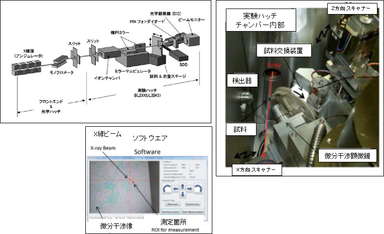
◆装置の特徴
・Sub-50nm集光システム(KBミラー)による高分解能マッピング・複数同時に元素マッピング情報を取得可能
・ズーム機能(ビームサイズの変更が可能)
・複数試料に対する自動測定が可能
・ユーザーフレンドリーなソフトウエアが完備
◆実験・試料準備
被測定物の形状、サイズ
・細胞内小分子:元素、薬剤、1原子ラベル脂質など代謝産物 (ホルマリン固定、瞬間凍結乾燥)
Scanning X-Ray Fluorescence Microscope
◆Equipment overview
This equipment uses nano-fluorescence X-ray analysis using X-ray nanobeams. High-sensitivity and high-resolution imaging of intracellular element distribution is possible.
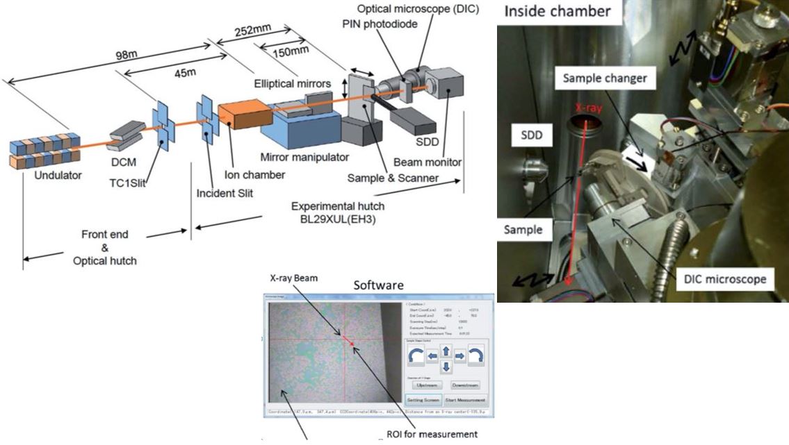
◆Features of the Equipment
・High-resolution mapping is possible with a sub-50nm focusing mirror (KB mirror).・Multiple mapping measurements can be acquired simultaneously.
・A zoom function exists (the beam size can be changed).
・It is possible to perform automatic measurements for multiple samples.
・The system is equipped with user-friendly software.
◆Experiment / sample preparation
Shape and size of the objects to be measured:
・Small molecules within the cell: metabolism products such as elements, medicines, and single atom label lipids (formalin fixation, instant freeze-drying).